It is quite difficult to imagine the world through other people's eyes, especially through the eyes of various animals. But new research using human retinas grown in the lab reveals that our vision is highly variable, even between different people.
And it may have something to do with how red and green cones are formed in our retinas. Cones are light-sensing cells in the eyes of vertebrates. The combination of their responses to different wavelengths makes color vision possible.
Humans and some related primates are the only mammals known to be able to see red as well as green and blue.
Other animals can also see red, like many birds and some insects. The type of vision possessed by animals is closely related to their evolution, along with the plants that produce fruits and flowers. This ability was very useful, for example, in finding ripe red apples in a dense green canopy.
Another outlier mammal with the ability to see red is the honey possum (Tarsipes rostratus). In an interesting example of convergent evolution, this Australian marsupial pollinator has a bird-like ability to forage for blush banksia nectar.
Red and green cones are essentially the same, but have slightly different chemistry that determines which colors they detect. Proteins called opsins come in two different “flavors”: red-sensitive or green-sensitive, and their genetic “recipes” sit side by side on her X chromosome.
Therefore, they are very easily confused during recombination, resulting in variations of congenital red-green color blindness.

Now, a new study sheds some light on what the key components that determine vision actually are, with only 4% variation between the genes encoding these proteins.
Although we previously thought that pyramidal cell decisions were essentially random, more recent studies have implicated thyroid levels.
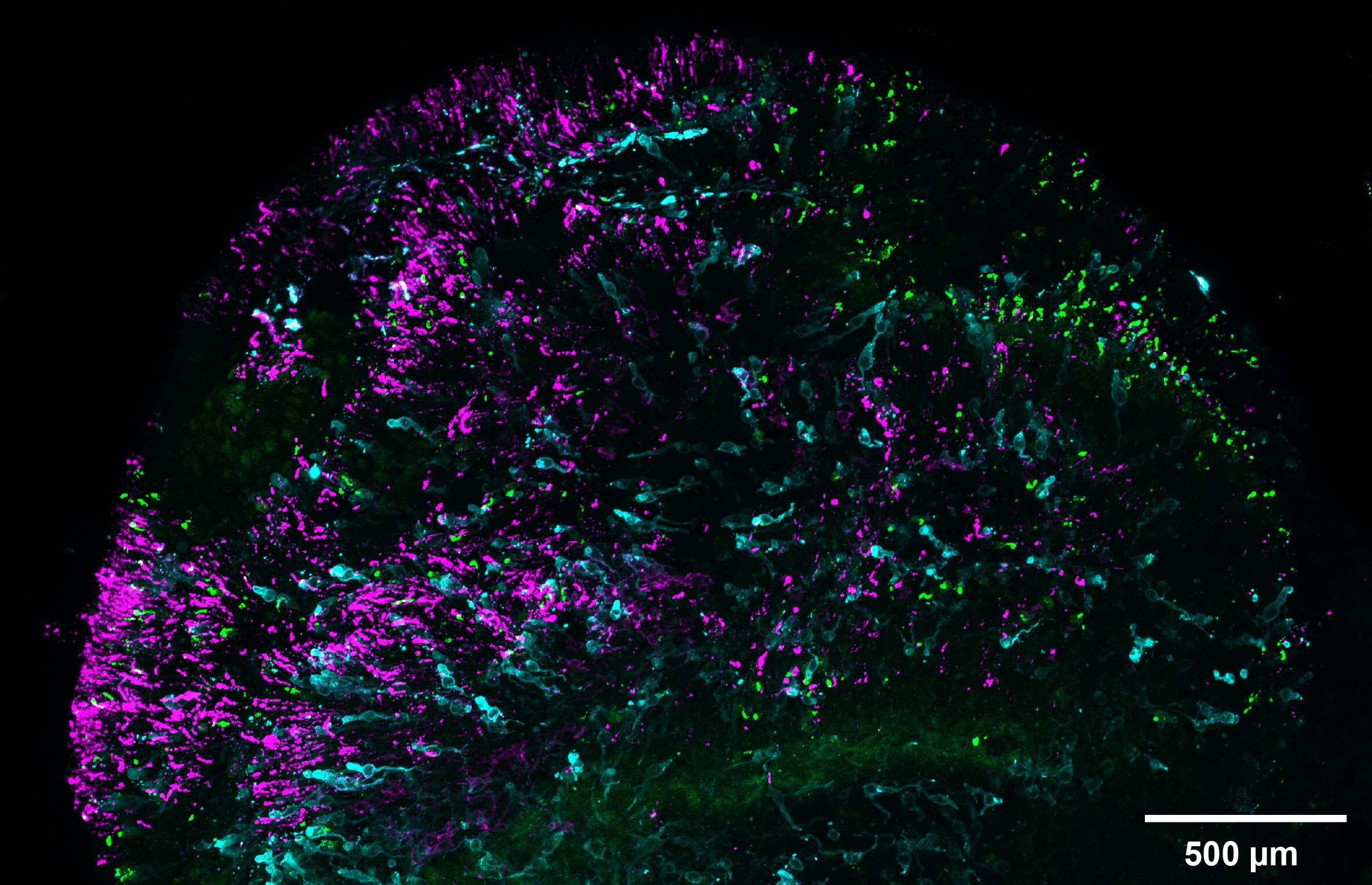
But a team of researchers from Johns Hopkins University and the University of Washington found that, at least in lab-grown retinas, levels of a vitamin A-derived molecule called retinoic acid govern the ratio of red-green cones. did.
“These retinal organoids allow us to study this very human trait for the first time,” said Robert Johnston, a developmental biologist at Johns Hopkins University. “It's a big question about what makes us human and what makes us different.”
In the laboratory, retinas exposed to more retinoic acid earlier in development (first 60 days) had a higher proportion of green cones throughout the organoids after 200 days, whereas immature retinas exposed to lower levels of acid The cones later developed into red cones.
Timing is also important. When retinoic acid was introduced after 130 days, the effect was the same as when nothing was added. This suggests that the acid determines the cone type early and that red cones cannot “switch” into already mature green cones.
Because all the retinas produced in the lab had similar cone densities, the researchers were able to rule out the possibility that cone cell death affected the red-to-green ratio.
Their findings have implications for understanding exactly how retinoic acid acts on genes, said developmental biologist Sarah Hadiniak, who co-authored the study while at Johns Hopkins University. It states that there is.
To find out how much this affects human vision, researchers studied the retinas of 738 adult men with no signs of color vision deficiency.
The researchers were surprised by the natural variation in the ratio of red to green cones across this group.
“Observing how the ratio of green to red cones changed in humans was one of the most surprising findings of the new study,” Hadiniak says.
It is unclear how such changes can occur without affecting visual acuity changes. As Johnston said, “If these cell types determine the length of human arms, the difference in proportions would result in strikingly different arm lengths.”
This research PLOS Biology.


